

CyTOF Helios & Hyperion
Helios analyzes individual cells labeled with stable metal isotopes using state-of-the-art inductively coupled plasma time-of-flight (TOF) technology. With 135 detection channels, Helios can simultaneously resolve multiple elemental probes at high acquisition rates, thereby maximizing the per-cell information obtained from a single sample.
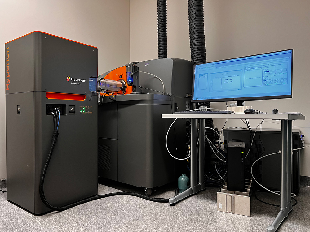
The Hyperion Imaging Systems combine a precisely directed laser beam focused to 1 μm to collect biological samples stained with metal-tagged Maxpar antibodies and direct these tags for analysis by CyTOF technology. The system provides spatial resolution and quantitation of up to 40 distinct markers on a single frozen or formalin-fixed, paraffin-embedded (FFPE) tissue sections.
Helios & Hyperion Location
Science Building, Room 533
Phone: 212-263-5907
Helios (Cell Suspension) Sample Preparation
- After staining, wash cells with Maxpar Cell Acquisition Solution Plus at least twice and leave cells pelleted at 2-8 ºC until ready to run.
- Resuspend the samples to the maximum recommended cell concentration of 1 x 10⁶ cells/mL with Maxpar CAS Plus containing 0.1X EQ4 beads immediately before data acquisition.
- Filter cells through 35 μm cell strainer cap tubes into a new 5 mL polypropylene tube.
Hyperion (Imaging Mode) Slide Preparation
- Follow the protocol to stain the slides.
- To save time, install CyTOF software on your own computer and make MCD file in advance, including slide image and detect template.
For more information about the related products and protocols, please check Standard BioTools website.